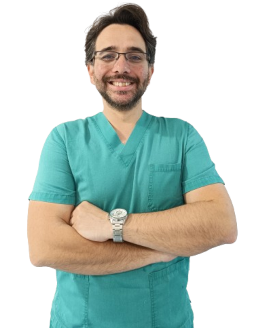
Sports Shoulder Surgery in Asturias
With 12 years of experience and advanced training, I specialize in innovative arthroscopic techniques to treat shoulder injuries in athletes. My personalized approach and commitment to excellence have established me as a leader in sports shoulder surgery in Asturias. I offer optimal results and a fast and effective recovery for all my patients, ensuring each treatment is tailored to individual needs. My goal is to return athletes to their maximum performance in the shortest possible time.

My Areas of Expertise
Shoulder Instability Repair: Arthroscopic Bankart
Shoulder Instability Repair: Arthroscopic Remplissage
Shoulder Instability Repair: Arthroscopic Bone-Block and Open Latarjet
Arthroscopic Rotator Cuff Repair
Tenodesis and tenotomy of the long head biceps
Subacromial decompression
Arthroscopic Superior Capsule Reconstruction
Subacromial Spacer Implantation
Subacromial Spacer Implantation
Minced Cartilage
Repair and Reconstruction of Acromioclavicular Dislocations
Glenoid, Proximal Humerus and Clavicle Fractures
My Shoulder Treatments in Detail
I have extensive experience and training in new techniques of complex shoulder surgery: arthroscopy, tendon, labrum and ligament repair, fracture fixation, and joint reconstruction. I am dedicated to excellence and provide individual care using the latest technology to get you back to function as soon as possible.
Shoulder instability occurs when the shoulder joint cannot be held in its normal position and repeatedly dislocates (pops out) or subluxates (partially dislocates). This may be due to trauma, repetitive overhead activities, or congenital conditions that weaken or stretch the ligaments, tendons, and muscles that hold the joint together.
Symptoms of shoulder instability include pain, a sense of failure, and repeated dislocation of the joint. Chronic instability can involve constant discomfort, weakness, and loss of function, making it difficult to perform daily activities and sports.
Non-surgical treatments are indicated in older patients with little overhead activity and when there has been no previous traumatic dislocation. In cases of traumatic dislocations, especially in young, athletic, or physically active patients, joint stabilization is the best treatment option.
Arthroscopic surgery is the ideal alternative in the vast majority of cases. Bankart repair is an arthroscopic procedure to repair glenoid labrum tears, a cartilaginous structure crucial for shoulder stability. It is especially effective for anterior instability caused by traumatic dislocations. Another option is the Remplissage, mainly used when there are bony defects in the humeral head, known as Hill-Sachs lesions. This is a complementary procedure to the Bankart repair in which the defect is filled with rotator cuff tissue to improve joint stability.
For cases with significant glenoid bone loss, Bone-Block is the best option. It consists of transferring a block of bone from the patient's pelvis (autograft) or from a donor tibia (allograft) into the glenoid cavity to increase the bone surface and provide more stability. Latarjet is an advanced surgical technique in which the coracoid and its adjacent tendons are transferred to the glenoid, achieving a triple effect of increased bone surface area, dynamic blocking, and reinforcement of the anterior shoulder capsule. This technique is usually performed open, as the arthroscopic approach is associated with a higher rate of complications.
Subacromial syndrome (subacromial impingement syndrome) is a condition in which the shoulder structures are compressed in the subacromial space. This space under the acromion houses the rotator cuff tendons and the subacromial bursa. When these are compressed, pain, swelling, and loss of function occur. Occasionally, this compression generates constant friction of the bone on the supraspinatus tendon, which can degenerate into calcific tendinitis or tendon rupture.
Typical symptoms of subacromial syndrome are pain that may radiate into the arm, especially during overhead activities and lying on the affected side, and may result in weakness of the shoulder muscles and loss of motion.
Initial treatment is usually conservative except in severe cases.
When conservative measures do not provide sufficient relief, arthroscopic subacromial decompression may be necessary. In this surgery, bone spurs and inflamed tissues within the subacromial space are removed. This creates more room for the rotator cuff tendons to reduce compression and relieve symptoms. If associated with calcifying tendinopathy, a small cut is also made in the tendon to drain the calcification, leaving it unrepaired if it is very small and repairing it if a larger one is needed. When there is significant rotator cuff damage, surgical repair of the torn tendons may be necessary to restore shoulder function and stability.
The rotator cuff is formed by the subscapularis, supraspinatus, infraspinatus, and teres minor tendons, and its function is to provide mobility and stability to the joint. Injuries may be due to acute trauma or degenerative changes due to repetitive overhead activities and age-related wear and tear.
Symptoms of rotator cuff injuries vary depending on severity but typically include persistent pain, especially with heavy lifting or overhead activities, and discomfort when sleeping on the affected side. They may also experience weakness, limited range of motion, and a crunching or popping sensation when moving the shoulder. In case of severe tears, daily activities such as dressing or combing hair may become difficult.
Treatment options for rotator cuff injuries depend on the extent of damage and the patient's functional needs. Nonsurgical treatments are reserved for older patients and those with a good range of motion and function.
Surgical treatment is indicated when conservative measures are insufficient, or if the injury is severe. The most common treatment is arthroscopic repair, which seeks to bring the tendons to the bone and fix them by tunneling through the humerus or using implants with sutures. The use of biological augmentation patches can help improve healing potential. In cases of non-repairable tears or failure of a previous repair, capsular reconstruction may be considered, which consists of using a graft to fill the defect and center the humerus in the glenoid. A tendon transfer may also be indicated, in which a tendon close to the shoulder is brought to the insertion area of the injured one. This surgery has a very harsh and prolonged postoperative period and is not the ideal option for most patients. Another very useful option in older patients without severe osteoarthritis is to implant a subacromial balloon, which centers the head of the humerus in the glenoid without needing to repair the supraspinatus tendon. If the tear is not repairable and there is already severe cartilage damage, the best alternative is implanting a prosthesis.
Long head biceps tendon injury, also known as biceps tendinopathy or biceps tendonitis, is an inflammation or damage to the tendon that runs from the biceps muscle over the top of the shoulder and attaches to the labrum. This type of injury is usually caused by repetitive overhead activities, acute trauma, or degenerative changes, and causes pain and functional disability in the shoulder. It is commonly associated with rotator cuff injuries, especially subscapularis.
Patients with long head of biceps injury have pain in the front of the shoulder that may radiate to the elbow. The pain is usually worse with overhead activities, lifting, or stretching. Loss of strength is also common.
Treatment of long head of biceps injury depends on the severity of the condition and the patient's activity level. Initial treatment is usually conservative.
Surgical intervention may be necessary when conservative treatment does not work or the tendon is badly damaged. There are two options for surgical treatment. In young, thin patients, tenodesis is preferred, which consists of cutting the tendon and attaching it to the humerus instead of the glenoid, shortening the lever arm. Its main advantage is aesthetics and, to a lesser extent, better strength, although only significant in professional pitchers and other jobs requiring great overhead power. Tenotomy is the ideal option in older patients, obese or with low functional demand, and is based on making a cut in the tendon inside the joint and not repairing it.
Cartilage injuries in the shoulder affect the smooth tissue that lines the ends of the bones within the joint. They may be due to acute trauma, repetitive stress, or degenerative diseases such as osteoarthritis. Damaged cartilage can cause pain, decreased function, and general shoulder instability.
Symptoms of shoulder cartilage injuries are persistent pain in the shoulder, especially during movement or weight-bearing activities. Patients often report feeling stiffness or locking of the joint, with swelling and stiffness. This can limit the range of motion and overall shoulder strength and impact daily activities and athletic performance.
Treatment of cartilage lesions consists of restoring the articular surface and relieving symptoms. A new technique involves using minced cartilage to repair the damaged area. This involves taking small pieces of healthy cartilage from the patient and transplanting them to the area of injury.
The procedure begins with an arthroscopic examination to assess the extent of cartilage damage. Healthy cartilage is removed from the edges of the defect and, if insufficient, from a non-weight-bearing, non-marring area of the joint. Upon removal, it is finely cut and mixed with a biological scaffold of platelet-rich plasma (PRP) that promotes cell adhesion and growth. This mixture is applied to the area of injury to create an environment conducive to cartilage regeneration and healing.
Minced cartilage has many advantages, such as minimally invasive surgery, shorter recovery time, and natural tissue regeneration. The biological scaffold favors the integration and growth of new cartilage cells to improve joint function and relieve pain, avoiding, or at least delaying, the need to consider joint replacement surgery (prosthesis).
Commitment to advanced surgical techniques and patient-centered care are key to ensuring that young patients with shoulder cartilage injuries receive the best treatment and return to their daily activities with improved joint function and quality of life.
Acromioclavicular (AC) instability is an injury in which the AC joint (where the clavicle attaches to the acromion of the scapula) is broken. It is usually caused by trauma, such as a fall or direct blow to the shoulder, which causes partial or total dislocation. Repetitive stress injuries and degenerative changes can also cause instability of the AC joint. It is classified into types I to VI.
The most frequent symptoms are localized pain in the joint, limitation of mobility (common to all), and deformity in lesions type III and onwards.
El tratamiento conservador se reserva para las lesiones tipo I y II, y las tipo III en pacientes con escasa demanda funcional por encima de la cabeza.
Surgical treatment is indicated in type IV, V, and VI lesions, and type III lesions in patients with intense work or sports activity. Although there are several types of treatment, nowadays two types of surgery are usually performed. Primary repair is performed in acute injuries (up to 1 month after the trauma). It consists of fixing the clavicle to the coracoid process (area of the scapula where the main ligaments are inserted) using a system formed by a suture of very high resistance in the form of thread or tape, joined by two small metal plates that rest on the surface of the bone house. It can be done openly by a minimal incision or arthroscopically assisted. This option is useful if other injuries are suspected within the joint, typically to the labrum (the structure that centers the round head of the humerus on the flat surface of the glenoid).
In case of chronic injuries (more than 1 month of evolution) or in patients who play high-contact sports (rugby, basketball, etc), reconstruction is the best alternative. This is performed using a tendon from the patient's knee or ankle (autograft) or from a donor (allograft), which is passed in a figure of 8 between the clavicle and the coronoid process, seeking to mimic the insertion of the injured ligaments. Although it is more stable, it has the added risk of using one's tissue, with the possible complications of the donor area, or tissue from a donor.
Las fracturas de glena, clavícula y húmero proximal son lesiones importantes del hombro, cada una con problemas particulares que requieren intervenciones específicas.
Glenoid fractures affect the flat part of the scapula that articulates with the ball of the humeral head. They almost always occur from high-energy trauma such as falls or traffic accidents. Symptoms are severe shoulder pain, swelling, bruising, limited range of motion, and a feeling of instability or snapping. Surgical treatment is indicated if the joint surface is involved and if associated with frank dislocation or instability. It consists of internal fixation, either open or arthroscopic, in which the bony fragments are realigned and fixed with screws or suture implants to ensure correct alignment and stability of the joint.
Clavicle fractures are usually caused by falls on the shoulder, direct blows, or traffic accidents. Patients usually present with acute pain at the fracture site, swelling, tenderness, and visible deformity that limits arm movement. Surgical intervention, especially in displaced, articular, or complex fractures, involves open reduction and internal fixation (ORIF) with a plate and screws to restore anatomic alignment, allow early mobilization and rehabilitation, prevent malunion, and improve functional outcomes.
Proximal humerus fractures occur in the shoulder joint area mainly after falls. They are more frequent in older adults due to osteoporosis. Symptoms are severe shoulder pain, swelling, bruising, limitation of arm motion, and sometimes a palpable gap at the fracture site. Treatment depends on the fracture pattern and patient characteristics. When surgery is indicated, ORIF is usually performed to realign and stabilize the fracture with a plate and screws. Occasionally, bone grafting is necessary to fill significant bone defects. In fractures where the sphericity of the humeral head cannot be restored or blood supply is compromised, partial (hemiarthroplasty, indicated in young patients with an intact rotator cuff) or total (reverse shoulder arthroplasty, indicated for older patients or those with non-repairable rotator cuff injuries) prosthesis implantation may be considered.